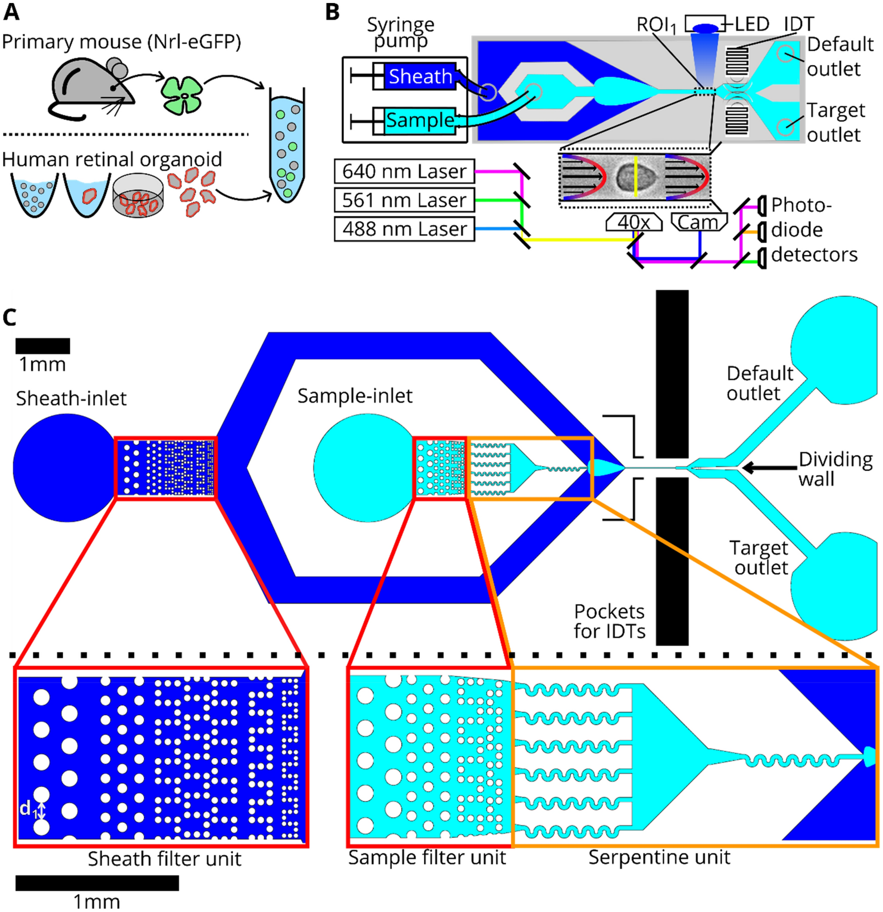
43 which label or labels indicate(s) the antigen binding site?
Different Ways to Add Fluorescent Labels - Thermo Fisher Scientific Labeling various targets with separate fluorescent colors allows you to visualize different structures or proteins within a cell in the same experiment. Ways to fluorescently label your target include fluorescent dyes, immunolabeling, and fluorescent fusion proteins —all of which can provide a means to selectively mark structures and proteins ... Overview of Protein Labeling | Thermo Fisher Scientific - US Overview of Protein Labeling. Biological research often requires the use of molecular labels that are covalently attached to a protein of interest to facilitate detection or purification of the labeled protein and/or its binding partners. Labeling strategies result in the covalent attachment of different molecules, including biotin, reporter ...
EIAs and ELISAs | Microbiology | | Course Hero A red color (from gold particles) or blue (from latex beads) developing at the test line indicates a positive test. If the color only develops at the control line, the test is negative. Like ELISA techniques, lateral flow tests take advantage of antibody sandwiches, providing sensitivity and specificity.

Which label or labels indicate(s) the antigen binding site?
Exam 2 Lab Flashcards | Chegg.com Which label or labels indicate (s) the antigen binding sight? The two sides labeled D The two sides labeled A The single location labeled B The two sites labeled C A What defense mechanism is shown in the images above? Complement Pyrogen Interferon Perforin Complement What defense mechanism is shown in this image? PDF Secondary - Novus Bio Variable domain heavy chain Contains antigen binding site, confers antibody specificity (H+L) Heavy + light Whole immunoglobulin (heavy and light chain) α Alpha heavy chain IgA class δ Delta heavy chain IgD class ε Epsilon heavy chain IgE class γ Gamma heavy chain IgG class µ Mu heavy chain IgM class κ Kappa light chain λ Lambda light chain Multiplex label-free biosensor for detection of ... - ScienceDirect The labels may destroy the binding sites that participate in antibody-antigen interactions, on the one hand, and non-specifically interact with proteins immobilized on a surface, on the other hand (Cooper, 2009; Fan et al., 2008; Orlov et al., 2018; Seydack, 2005). The label-free techniques are not prone to the label-associated issues.
Which label or labels indicate(s) the antigen binding site?. Anatomy Labs for Exam 2 Flashcards | Quizlet Which label or labels indicate(s) the antigen binding site? The two sites labeled with the letter A. What defense mechanism is shown in the images above? Complement (group of proteins that work together, binding to antibodies and forming large holes in target cells) What defense mechanism is shown in this image? Destruction by an NK cell. Anatomy and Physiology 2: Chapter 22: Immune System Indicate whether the label identifies a specific or nonspecific form of defense. Specific: B-lymphocytes, antibodies, agglutinin, complement (antibody-dependent pathway), major histocompatability complexes, cytotoxic T-cells, memory T-cells, plasma cells, immunoglobulin, T-lymphocytes, antigen presenting cells, helper T-cells, CD4+ cells Structure of Antibody (With Diagram) | Organisms - Biology This variable region, composed of 110-130 amino acids, gives the antibody its specificity for binding antigen. ADVERTISEMENTS: Antibodies are of five classes - IgG, IgA, IgM, IgD and IgE. Ig stands for immunoglobulins. IgG constitutes to about 75% of the total antibodies. IgE is involved in allergy and IgM is formed during the primary response. Exam 3: Mastering Lymphatic System (#6) Flashcards | Quizlet Which of these statements about lymphocytes is false? -They bind antigens. -They mostly occur in lymphoid tissues. -They are phagocytic. -They occur as B, T, and NK types. they are phagocytic Classes of lymphocytes T cells B cells NK cells T cells are approximately ___% of circulating lymphocytes. 80% B cells are ___% of circulating lymphocytes.
HW 29.docx - Best of Home Work (Exercise 29: Blood) Drag... Label the red blood cells with the correct antigen (s). Left to right… a antigen, b antigen, a & b, none Left to right … a antigen , b antigen , a & b , none Antibodies are proteins that have a lock-and-key recognition for their antigen established by the antigen-binding site on the antibody. › 37006818 › Junqueiras_BasicJunqueira's Basic Histology Text and Atlas, 14th Edition There is shortage of references in higher teaching institutions especially in newly opened institutions engaged in training of various Veterinary professionals in the country. Lateral flow assays: Principles, designs and labels An alternative biorecognition molecule of antibody is immunogen, which is a cheaper biomolecule than an antibody, since the production cost of antibody is high. The binding of antibody to colloidal gold occurs by means of hydrophobic residues in the antigen-binding site of antibody. This way, the binding sites of an antibody are decreased. Top 7 Types of Immunochemical Techniques Used in Biochemistry Technique # 1. Immunoassay: An immunoassay is a biochemical test that measures the concentration of a substance in a biological liquid, typically serum or urine, using the reaction of an antibody or antibodies to its antigen. The assay takes advantage of the specific binding of an antibody to its antigen. Monoclonal antibodies are often used as ...
A&P2 Lab 10 HW Flashcards | Quizlet Study with Quizlet and memorize flashcards containing terms like Drag the labels onto the diagram to identify the lymphoid tissues and organs of the lymphatic system., Drag the labels onto the diagram to identify the structural features of the spleen., Which of the labels indicates a structure through which lymph flows? and more. dailymed.nlm.nih.gov › dailymed › drugInfoDailyMed - TRELEGY ELLIPTA- fluticasone furoate, umeclidinium ... May 11, 2022 · In vitro plasma protein binding in human plasma was on average 89%. Vilanterol: Following intravenous administration to healthy subjects, the mean volume of distribution at steady state was 165 L. In vitro plasma protein binding in human plasma was on average 94%. Elimination Immunolabeling | Thermo Fisher Scientific - US For direct immunofluorescence, the antibody binding the epitope is labeled with fluorophores (green). For indirect or secondary detection, the primary antibody binds the epitope and a fluorophore-labeled secondary antibody (purple) that has specificity for the primary antibody binds to it. Direct immunofluorescence Real-Time, label-free monitoring of tumor antigen and serum antibody ... The binding kinetics and affinity values of anti-NY-ESO-1 monoclonal antibody, ES121, to the cancer-testis antigen NY-ESO-1 were determined according to the surface heterogeneity model and resulted in K D values of 1.3×10 −9 and 2.1×10 −10 M. The reconfigured instrument was then used to measure the interaction between tumor antigens and ...
High-Affinity Points of Interaction on Antibody Allow ...
M A&P Introduction to Lymphatic System and Immunity Label the cause of infection and some structures involved in fighting the infection. After the proteins are separated by electrophoresis, the _____. ... Which label or labels indicate(s) the antigen binding site? the two sites labeled with the letter A. Steps in antigen presentation include which of these?

Double and triple immunostaining using secondary antibodies
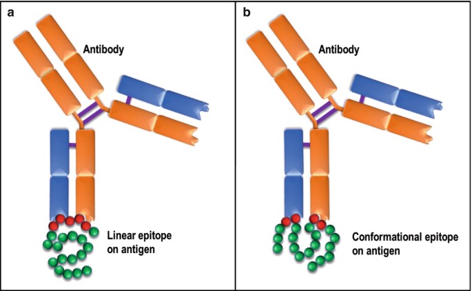
Antibody Types: IgM, IgA, IgD, IgG, IgE and Camelid Antibodies However, since pentameric IgM has 10 antigen binding sites, it has higher avidity (overall binding strength) for antigens than IgG and acts as an excellent activator of the complement system and ...

Phenotypic determinism and stochasticity in antibody ...
Multiplex label-free biosensor for detection of ... - ScienceDirect The labels may destroy the binding sites that participate in antibody-antigen interactions, on the one hand, and non-specifically interact with proteins immobilized on a surface, on the other hand (Cooper, 2009; Fan et al., 2008; Orlov et al., 2018; Seydack, 2005). The label-free techniques are not prone to the label-associated issues.
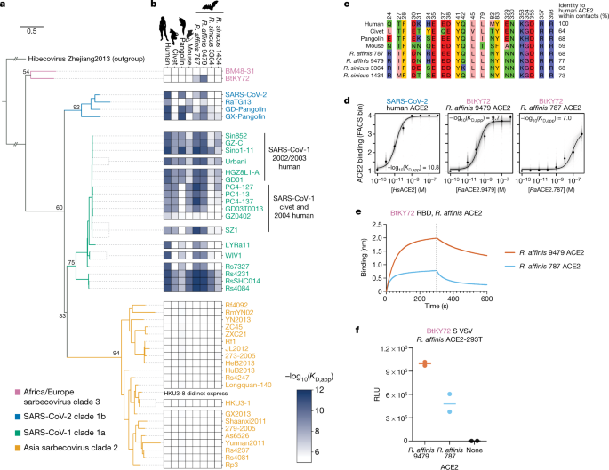
ACE2 binding is an ancestral and evolvable trait of ...
PDF Secondary - Novus Bio Variable domain heavy chain Contains antigen binding site, confers antibody specificity (H+L) Heavy + light Whole immunoglobulin (heavy and light chain) α Alpha heavy chain IgA class δ Delta heavy chain IgD class ε Epsilon heavy chain IgE class γ Gamma heavy chain IgG class µ Mu heavy chain IgM class κ Kappa light chain λ Lambda light chain
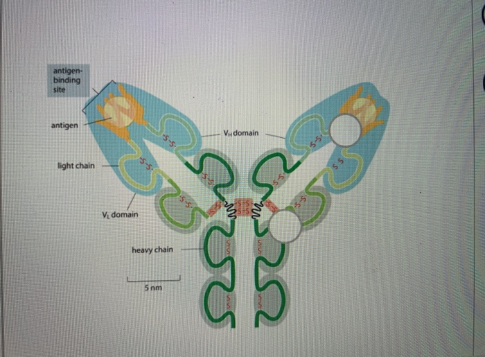
Label-free imaging flow cytometry for analysis and sorting of ...
Exam 2 Lab Flashcards | Chegg.com Which label or labels indicate (s) the antigen binding sight? The two sides labeled D The two sides labeled A The single location labeled B The two sites labeled C A What defense mechanism is shown in the images above? Complement Pyrogen Interferon Perforin Complement What defense mechanism is shown in this image?

Deciphering spatial protein-protein interactions in brain ...

Stable Isotope Labeling of Amino Acids in Flies (SILAF ...

Analysis of Cell-Surface Receptor Dynamics through Covalent ...
Solved within the representation of an antibody, identify ...

Why Site-Specific — AlphaThera
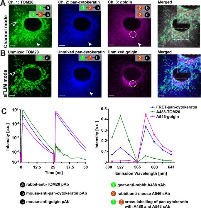
Multi-target immunofluorescence by separation of antibody ...

New Insights into the DT40 B Cell Receptor Cluster Using a ...

Affinity-Based Methods for Site-Specific Conjugation of ...

Ch. 16- Prep for Exam Flashcards | Quizlet
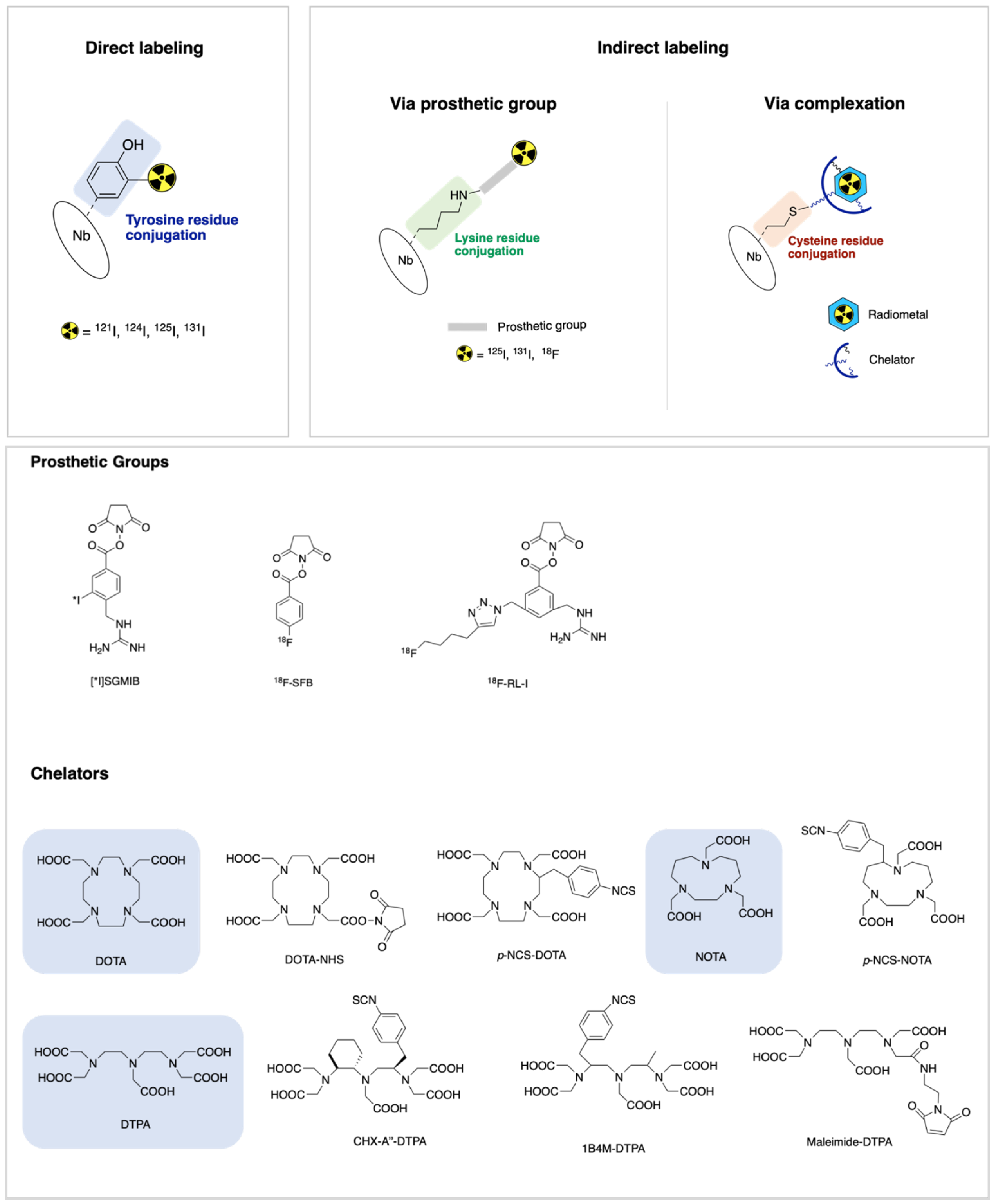
IJMS | Free Full-Text | Nanobody-Based Theranostic Agents for ...

Multicolor labeling of HA-tagged proteins in vivo. a ...

Systematic identification of engineered methionines and ...

Site-Specific Antibody Labeling with oYo-Link

A&P2 Lab 10 HW Flashcards | Quizlet
Mapping Protein Binding Sites and Conformational Epitopes ...
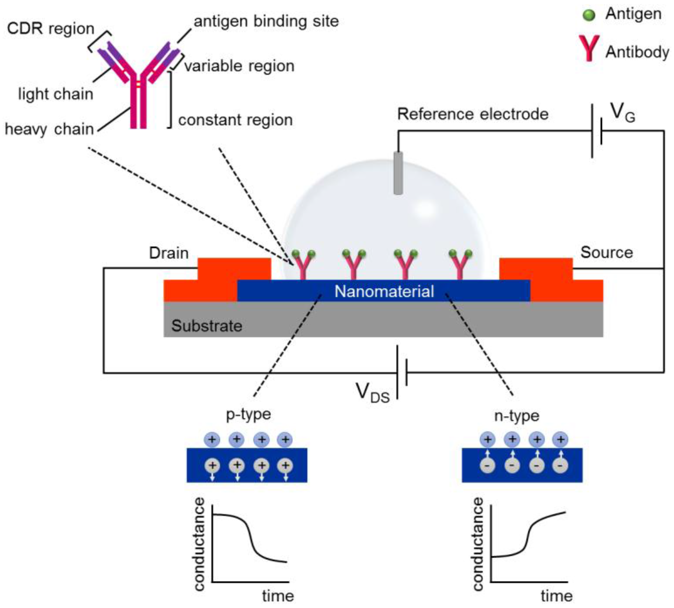
Chemosensors | Free Full-Text | Recent Trends in Field-Effect ...

Week 11: Blood Typing Flashcards | Quizlet

Electric-Field-Mediated In-Sensor Alignment of Antibody's ...

Antibody Labeling - an overview | ScienceDirect Topics
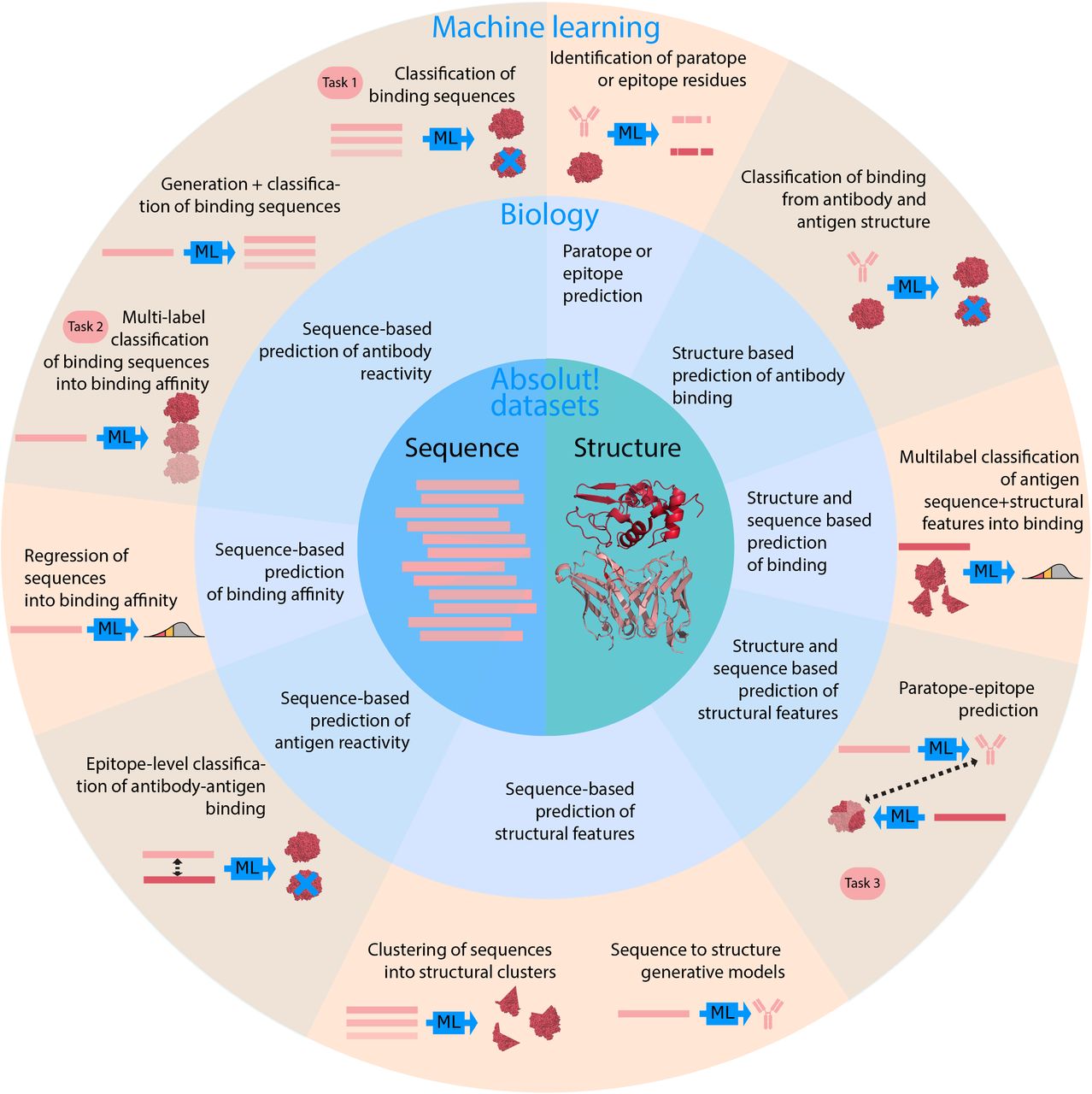
One billion synthetic 3D-antibody-antigen complexes enable ...

Why Site-Specific — AlphaThera

One billion synthetic 3D-antibody-antigen complexes enable ...

Deep Mutational Scanning of SARS-CoV-2 Receptor Binding ...

Labeling of ORAI1 proteins in the plasma membrane of HEK ...

Cell cycle labelling. Schematic representation of the cell ...
Using the antibody-antigen binding interface to train image ...

Affinity-Based Methods for Site-Specific Conjugation of ...

A&P2 Lab 13 HW, A&P2 Lab 12 HW, A&P2 Lab 11 HW, A&P2 Lab 10 ...

Antibody Structure

Antibody Structure and Function | Sino Biological
Immunoassays | SpringerLink

AzG is compatible with metabolic RNA labeling in E. coli. (A ...

One billion synthetic 3D-antibody-antigen complexes enable ...

Why Site-Specific — AlphaThera
Bioorthogonal labeling of transmembrane proteins with non ...

Draw a neat labeled diagram of an antibody molecule and ...

Small Molecule Interactome Mapping by Photoaffinity Labeling ...

Ch. 16- Prep for Exam Flashcards | Quizlet
Komentar
Posting Komentar